
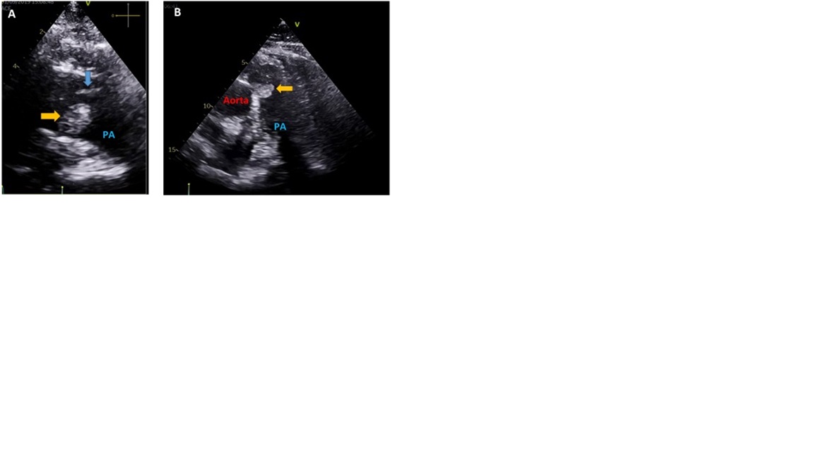
Papillary Fibroelastoma on the Pulmonary Valve: An Uncommon and Unexpected Diagnosis
DOI:
https://doi.org/10.24950/rspmi.634Keywords:
Cardiac Papillary Fibroelastoma, Echocardiography, Heart Neoplasms/diagnostic imaging, Pulmonary Valve/diagnostic imagingDownloads
References
Gowda RM, Khan IA, Nair CK, Mehta NJ, Vasavada BC, Sacchi TJ. Cardiac papillary fibroelastoma: a comprehensive analysis of 725 cases. Am Heart J. 2003;146:404-10. doi: 10.1016/S0002-8703(03)00249-7
Rudkovskaia AA, Bandyopadhyay D. Intraluminal arterial filling defects misdiagnosed as pulmonary emboli. Clin Chest Med. 2018;39:505-13. doi:10.1016/j.ccm.2018.04.004
Palaskas N, Thompson K, Gladish G, Agha AM, Hassan S, Iliescu C, et al. Evaluation and Management of Cardiac Tumors. Curr Treat Options Cardiovasc Med. 2018;20:29. doi: 10.1007/s11936-018-0625-z.
Xi XY, Gao W, Gong JN, Guo XJ, Wu JY, Yang YH, et al. Value of 18F-FDG PET/CT in differentiating malignancy of pulmonary artery from pulmonary thromboembolism: a cohort study and literature review. Int J Cardiovasc Imaging. 2019;35:1395-403. doi: 10.1007/s10554-019-01553-5.
Konstantinides SV, Meyer G, Becattini C, Bueno H, Geersing GJ, et al; ESC Scientific Document Group. 2019 ESC Guidelines for the diagnosis and management of acute pulmonary embolism developed in collaboration with the European Respiratory Society (ERS). Eur Heart J. 2020;41:543-603. doi: 10.1093/eurheartj/ehz405.
Downloads
Published
How to Cite
Issue
Section
Categories
License
Copyright (c) 2022 Medicina Interna

This work is licensed under a Creative Commons Attribution-NonCommercial 4.0 International License.